Facts about the 3D X-ray

Definition:
Three-dimensional X-ray procedure using digital volume tomography (DVT)
Areas of application:
Mainly oral implantology, wisdom tooth surgery, bone augmentations, unclear complaints
Examination duration:
5 to 15 seconds (pure recording time)
X-rays play an important role in dental diagnostics. A two- or three-dimensional X-ray can be used to monitor the progress of dental treatment and to diagnose various diseases or malocclusions. Most x-rays are taken in two dimensions, but only show the teeth from the front. Although this may be sufficient, the possibilities of this diagnostic method are quickly exhausted. At our dental practice in Zurich, we therefore use 3D X-rays to take a detailed look at the bony structures of the jaw, which enables us to make a reliable diagnosis and check the situation.
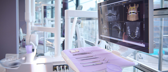
3D images provide us with details and therefore the greatest possible certainty during diagnosis and treatment. Similar to a CT scan, our X-ray machine allows us to take three-dimensional images of the jawbone and teeth with only a fraction of the radiation exposure. We can view and analyse the high-resolution images on our computer immediately after the uncomplicated imaging. In our dental practice in Zurich, we use this technology for various dental issues.
Among other things, 3D X-rays offer us the following options
- Precise planning and simulation of the positioning of dental implants
- Reliable assessment of the condition and state of the jawbone
- Assessment of the exact position of wisdom teeth
- Assessment of the preservability of diseased teeth
- Visualisation of inflammatory processes in the jawbone or maxillary sinuses
- View of the exact course of root canals
- Visualisation of the periodontium as a whole
- Diagnosis of diseases of the maxillary sinus
- Determination of the number of existing root canals
- Traceability of changes in the jawbone (e.g. cysts)
Frequently asked questions about 3D X-rays
When is a three-dimensional X-ray useful?
A three-dimensional X-ray is always useful when detailed images of the jaw and teeth are required (visualisation of the hard tissue). If an implantation is to be planned with millimetre precision, for example, we use digital volume tomography to produce an image of your jaw. With the 3D X-ray, we can precisely record bone and tissue structures and also analyse the position of wisdom teeth. This allows us to recognise sensitive structures, such as nerves, even before the actual treatment. The 3D X-ray is also used for unclear symptoms, as it often reveals hidden inflammation.
Does a 3D X-ray harbour risks?
There is only a low risk of radiation during an X-ray. We therefore take strict radiation protection measures in our practice to protect you and our staff. With 3D X-ray diagnostics, the overall radiation exposure is also very low.
What radiation am I exposed to during the 3D X-ray scan?
Compared to other procedures, digital volume tomography (DVT) is very low in radiation. Radiation exposure of around 50 to 150 microsieverts can be assumed for an image. By comparison, a computer tomography (CT) scan leads to an exposure of around 500 to 1500 microsieverts.
Does health insurance cover the costs of 3D X-rays?
As a rule, the costs of digital volume tomography are not covered by statutory health insurance. If you have private supplementary insurance, the costs may be covered. We therefore recommend that you consult with your supplementary insurance provider before the examination.
Your Benefits of Treatments at Our Dental Practice
- Owner-managed practice
- Central location near Zurich Main Station
- Over 20 years of experience in dentistry
- Dentists for implants, aligners, and reconstructive dentistry
- All treatments performed on-site
- Easy appointment scheduling & short waiting times
- Pain-free & gentle treatment methods
- Guaranteed quality according to SSO & ITI guidelines
- Warranty on dental and technical work in accordance with SSO standards
- Transparent, fair billing with optional flat-rate offers
Procedure for a three-dimensional X-ray scan
At our dental practice in Zurich, the comfort and safety of our patients is our top priority. For this reason, the 3D X-ray in our practice takes place in a specially equipped room. You can usually stand or sit in our X-ray room during the entertaining examination. Our competent staff will accompany you during the entire scan and explain the procedure to you in detail. You are also welcome to express your concerns about the examination so that we can take care of your well-being.
Before the examination, we will put a radiation protection waistcoat on you to minimise the radiation exposure to your body. You will then stand or sit in front of the X-ray machine, where our staff will position your head and align the machine. Once you have been optimally positioned, we will start the X-ray scan. For your own safety, our staff will have to leave the room for a short time, but will be available for you at all times. The pure exposure time is only 5 to 15 seconds.
During the scan, the 3D X-ray machine moves once around your head without you having to move. There is no direct contact and the examination is completely painless. During the short exposure time, the X-ray machine takes around 500 individual images of your jaw and teeth. These are available to us on our computer for analysis immediately after the X-ray. If you wish, we can discuss the images immediately afterwards or at a separate appointment.
Contact
Dentist Zurich - finest smile
Seidengasse 20
8001 Zurich
Opening hours:
Monday to Friday
7:00 am to 8:00 pm
Phone
044 504 57 56